A Hypothetical Model of
“Hormonal Multi-Resistance”
Hormones such as AVP and leptin, along with other mediators, can spontaneously induce PGE2 production, possibly as part of a homeostatic negative-feedback mechanism, since PGE2 has been reported to antagonize their effects (while they continue to stimulate its production). Excessive upregulation by additional factors may lead to a state of “PGE2 dominance.”
Its receptor EP3 plays important homeostatic roles. In individuals with EP3 overexpression (“EP3 up”) or underexpression (“EP3 down”), homeostasis may become disrupted. In this context, PGE2 may become a persistent limiting factor, establishing a self-reinforcing cycle of “Hormonal Multi-Resistance” that sustains chronically elevated PGE2 levels
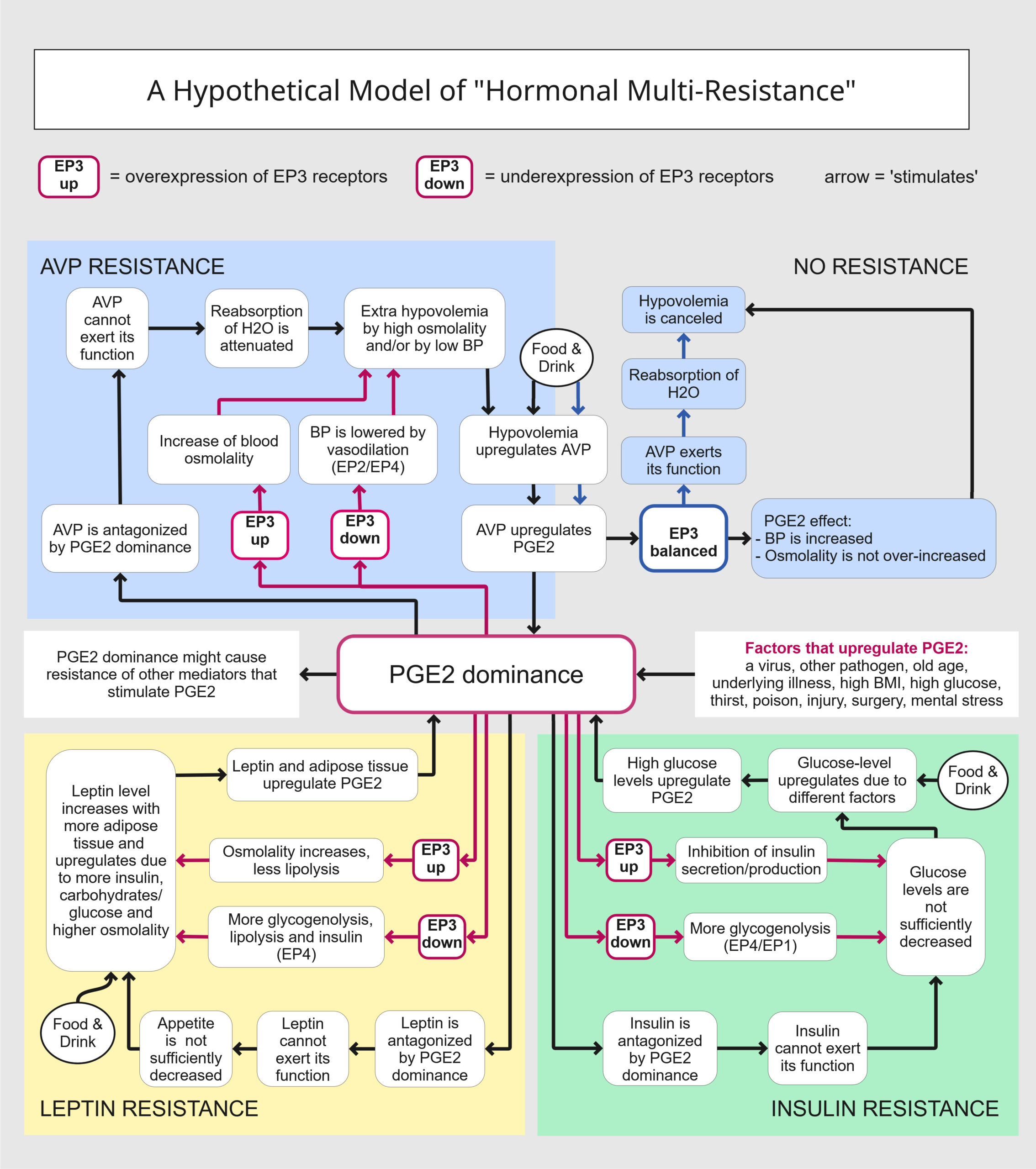
Clarification of hypothetical hormonal resistance loops
According to our hypothesis, PGE2 can antagonize the receptor binding of AVP, leptin, and insulin (while they presumably drive PGE2 upregulation as a limiter on their activity). These interdependent relationships may form vicious cycles – resistance loops – that potentially reinforce one another 1. We refer to this presumed phenomenon as ‘Hormonal multi-resistance”, illustrated in Figure 1.
A combination of factors may cause such a strong upregulation of PGE2 that its levels begin to dominate over certain endocrine hormones that spontaneously stimulate PGE2 synthesis but can also be antagonized by it. In this state of “PGE2 dominance”, the functions of arginine vasopressin (AVP), insulin, and/or leptin may be suppressed. Given the functions of the PGE2 receptor EP3, homeostasis can become chronically disrupted in individuals with either “EP3 up” or “EP3 down” (enhanced or insufficient EP3 capacity), driving upregulation of PGE2. This, in turn, promotes resistance to hormones that are antagonized by PGE2 but stimulate PGE2, ultimately reinforcing resistance to additional hormones – a process we describe as “Hormonal multi-resistance”.
-
-
AVP
-
When sufficiently upregulated, PGE2 has been reported to antagonize the activity of arginine vasopressin (AVP) 2. AVP, in turn, stimulates PGE2 production via its receptor V1 3. PGE2 increases osmolality through EP3 signaling 4 and lowers blood pressure through EP4 and EP2 signaling 5. Thus, PGE2 could cause AVP resistance in the following way: AVP is stimulated by hypovolemia when osmolality is high or when blood pressure is low. Hypovolemia induces AVP upregulation. However, under certain conditions, elevated PGE2 may dominate AVP activity, a state we term “PGE2 dominance”. PGE2 may prevent AVP from binding to its receptors by stimulating autoantibodies against AVP, as observed in another context 6. Polyuria resulting from AVP malfunction can cause low blood pressure and high osmolality. In this situation, increasing AVP levels cannot restore blood pressure or osmolality. Homeostatic imbalances will continue to stimulate AVP and PGE2 levels. Moreover, AVP itself stimulates PGE2, which, due to imbalanced EP3 expression, can either raise osmolality or lower blood pressure. Thus, both AVP and PGE2 may remain upregulated in a vicious loop.
-
-
Insulin
-
PGE2 can increase blood glucose by inhibiting insulin secretion through EP3 signaling 7, but it can also lower blood glucose by attenuating glycogenolysis through EP3 8. Either insufficient or enhanced EP3 capacity could lead to elevated glucose levels when stress factors upregulate PGE2. Elevated glucose levels, in turn, upregulate PGE2 9. We propose that PGE2 antagonizes insulin receptor binding by stimulating phosphorylation of insulin receptors (See section 5). When insulin cannot exert its function, glucose levels remain high, which in turn sustains PGE2 upregulation. In this way, glucose levels could remain elevated in both category A (EP3-up) and category B (“EP3 down”) individuals, albeit through different pathways, while PGE2 continues to antagonize insulin activity.
-
-
Leptin
-
As discussed in the previous section, either insufficient or enhanced EP3 signaling may lead to elevated glucose levels. Leptin, which is produced in adipose tissue, is upregulated by glucose and carbohydrate intake. Leptin has been repeatedly reported to stimulate PGE2 10. When PGE2 becomes sufficiently upregulated by various factors, it may antagonize leptin activity. Under conditions of PGE2 dominance, leptin receptor binding is impaired, resulting in leptin insensitivity. This leads to increased carbohydrate intake and adipose tissue accumulation, which, under inflammatory conditions, further stimulate PGE2 production. Chronically elevated glucose levels perpetuate leptin upregulation, but leptin remains functionally antagonized because high osmolality, adipose tissue, and leptin itself all maintain elevated PGE2 levels that block leptin activity.
According to our hypothesis, in addition to AVP, insulin, and leptin, other PGE-stimulating hormones and agents may exhibit markedly reduced responsiveness under conditions of PGE2 dominance.
Examples are:
-
-
Fibroblast growth factor 23 (FGF23)
-
One of the functions of fibroblast growth factor 23 (FGF23) is to promote phosphate excretion. Elevated phosphate levels have been reported in patients with long COVID patients 11. Inflammation has been shown to cause FGF23 resistance 12, and FGF23 itself stimulates PGE2 13. Elevated PGE2 could impair FGF23 activity, for example, by stimulating autoantibodies against FGF23, as observed in other contexts 14. Increased phosphate levels stimulate higher FGF23 concentrations, and FGF23 continues to upregulate PGE2, which in turn antagonizes its function. This may result in sustained elevation of phosphate, FGF23, and PGE2 levels.
-
-
Cytokine interferon-gamma (IFN-γ)
-
Patients who develop long COVID often show marked upregulation of specific cytokines, including interferon-gamma (IFN-γ) 15. Elevated IFN-γ can have detrimental effects 16, and IFN-γ resistance has been reported in other contexts 17. IFN-γ stimulates COX-2/PGE2 production 18. According to our hypothesis, sufficiently high PGE2 levels may antagonize IFN-γ activity, keeping IFN-γ chronically elevated while PGE2 remains upregulated, possibly by stimulating autoantibodies. Autoantibodies against IFN-γ that block receptor binding have indeed been observed in other contexts 19,20.
Referenties
1 Morrison, C. D., Huypens, P., Stewart, L. K. & Gettys, T. W. Implications of crosstalk between leptin and insulin signaling during the development of diet-induced obesity. Biochimica et Biophysica Acta (BBA) - Molecular Basis of Disease 1792, 409-416 (2009). https://doi.org/https://doi.org/10.1016/j.bbadis.2008.09.005
2 Hébert, R. L., Jacobson, H. R. & Breyer, M. D. PGE2 inhibits AVP-induced water flow in cortical collecting ducts by protein kinase C activation. Am J Physiol 259, F318-325 (1990). https://doi.org/10.1152/ajprenal.1990.259.2.F318
3 Wuthrich, R. P. & Vallotton, M. B. Prostaglandin E2 and cyclic AMP response to vasopressin in renal medullary tubular cells. Am J Physiol 251, F499-505 (1986). https://doi.org/10.1152/ajprenal.1986.251.3.F499
4 Hassouneh, R. et al. PGE2 receptor EP3 inhibits water reabsorption and contributes to polyuria and kidney injury in a streptozotocin-induced mouse model of diabetes. Diabetologia 59, 1318-1328 (2016). https://doi.org/10.1007/s00125-016-3916-5
5 Wang, L., Wu, Y., Jia, Z., Yu, J. & Huang, S. Roles of EP Receptors in the Regulation of Fluid Balance and Blood Pressure. Front Endocrinol (Lausanne) 13, 875425 (2022). https://doi.org/10.3389/fendo.2022.875425
6 De Bellis, A. et al. Detection of vasopressin cell antibodies in some patients with autoimmune endocrine diseases without overt diabetes insipidus. Clin Endocrinol (Oxf) 40, 173-177 (1994). https://doi.org/10.1111/j.1365-2265.1994.tb02464.x
7 Kimple, M. E. et al. Prostaglandin E2 receptor, EP3, is induced in diabetic islets and negatively regulates glucose- and hormone-stimulated insulin secretion. Diabetes 62, 1904-1912 (2013). https://doi.org/10.2337/db12-0769
8 Püschel, G. P., Kirchner, C., Schröder, A. & Jungermann, K. Glycogenolytic and antiglycogenolytic prostaglandin E2 actions in rat hepatocytes are mediated via different signalling pathways. Eur J Biochem 218, 1083-1089 (1993). https://doi.org/10.1111/j.1432-1033.1993.tb18468.x
9 Sitter, T. et al. High glucose increases prostaglandin E2 synthesis in human peritoneal mesothelial cells: role of hyperosmolarity. J Am Soc Nephrol 9, 2005-2012 (1998). https://doi.org/10.1681/asn.V9112005
10 Ruiz-Heiland, G., Yong, J. W., von Bremen, J. & Ruf, S. Leptin reduces in vitro cementoblast mineralization and survival as well as induces PGE2 release by ERK1/2 commitment. Clin Oral Investig 25, 1933-1944 (2021). https://doi.org/10.1007/s00784-020-03501-3
11 Corrêa, H. L. et al. Phosphate and IL-10 concentration as predictors of long-covid in hemodialysis patients: A Brazilian study. Frontiers in Immunology 13 (2022). https://doi.org/10.3389/fimmu.2022.1006076
12 Rodríguez-Ortiz, M. E. et al. Inflammation both increases and causes resistance to FGF23 in normal and uremic rats. Clin Sci (Lond) 134, 15-32 (2020). https://doi.org/10.1042/cs20190779
13 Syal, A. et al. Fibroblast growth factor-23 increases mouse PGE2 production in vivo and in vitro. Am J Physiol Renal Physiol 290, F450-455 (2006). https://doi.org/10.1152/ajprenal.00234.2005
14 Roberts, M. S. et al. Autoimmune hyperphosphatemic tumoral calcinosis in a patient with FGF23 autoantibodies. J Clin Invest 128, 5368-5373 (2018). https://doi.org/10.1172/jci122004
15 Krishna, B. A. et al. Spontaneous, persistent, T cell-dependent IFN-γ release in patients who progress to Long Covid. Sci Adv 10, eadi9379 (2024). https://doi.org/10.1126/sciadv.adi9379
16 Kak, G., Raza, M. & Tiwari, B. K. Interferon-gamma (IFN-γ): Exploring its implications in infectious diseases. Biomol Concepts 9, 64-79 (2018). https://doi.org/10.1515/bmc-2018-0007
17 Fojtova, M. et al. Development of IFN-γ resistance is associated with attenuation of SOCS genes induction and constitutive expression of SOCS 3 in melanoma cells. British Journal of Cancer 97, 231-237 (2007). https://doi.org/10.1038/sj.bjc.6603849
18 Kawada, N. et al. Interferon gamma stimulates prostaglandin E2 production by mouse Kupffer cells. Prostaglandins Leukot Essent Fatty Acids 40, 275-279 (1990). https://doi.org/10.1016/0952-3278(90)90049-q
19 Chawansuntati, K., Rattanathammethee, K. & Wipasa, J. Minireview: Insights into anti-interferon-γ autoantibodies. Exp Biol Med (Maywood) 246, 790-795 (2021). https://doi.org/10.1177/1535370220981579
20 Shih, H. P. et al. Pathogenic autoantibodies to IFN-γ act through the impedance of receptor assembly and Fc-mediated response. J Exp Med 219 (2022). https://doi.org/10.1084/jem.20212126